Outpatient Spine Surgery Mishawaka
Surgical Procedures
Our highly trained surgeon, Dr. Jamie Gottlieb, will have a thorough conversation about all our treatment options. Surgery is not recommended for every patient and usually is reserved for patients that have exhausted all other forms of treatment. If non-surgical treatments have failed or not progressed as planned, surgery can be arranged with Dr. Gottlieb and his team.
Procedure Information Packets
Some of our most common procedures for outpatient spine surgery in Mishawaka are listed below. Click the procedure name to open a PDF document in a new window.
 Anterior Cervical Discectomy and Fusion
Overview
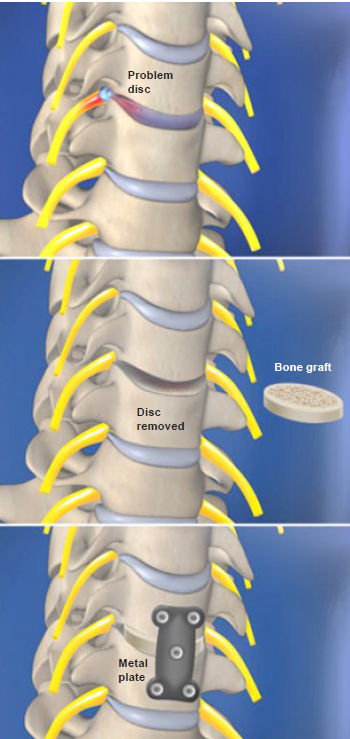
This surgery is where the disc(s) in the cervical region (neck) are removed which will release the pressure on the nerves in the spine. The disc space is then filled with bone graft which will help the vertebrae grow into one solid unit. This fusion will help keep the shape and form as well as provide space and room for the nerve roots and spinal cord.
Procedure
Incision Created
The surgeon performs this procedure through an incision on the front of the neck.
Disc Removed
The diseased or damaged disc is removed. As pressure is removed from the pinched nerve roots, pain is relieved.
Graft Inserted
The space above and below the removed disc is cleared and prepared for a bone graft. The graft is placed between the vertebrae.
Metal Plate Attached
The surgeon may screw a small metal plate over the area to hold the bones in place while the vertebrae heal.
End of Procedure
During the healing process, the bone graft knits together with the vertebrae above and below to form a new bone mass called a fusion.
Conditions Treated with Anterior Cervical Discectomy and Fusion
Spinal Stenosis
Rupture Disc
Pinched Nerve
Estimated Length of Stay in Hospital
The usual length of stay in the hospital for decompression and spine fusion surgery varies from one to three days. More than likely a cervical collar will be worn post-operatively.
Laminectomy
Overview
This procedure is performed through an incision on the lower back. The surgeon removes a section of bone, called the lamina, from one or more vertebrae. This relieves pressure on the nerve roots caused by stenosis (a narrowing of the spinal canal).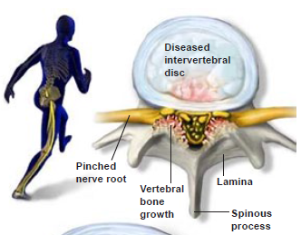
Procedure
First, the surgeon removes the spinous process (the portion of the vertebra that protrudes
furthest from the back of the spine). These are the bones that you feel when you touch the
middle portion of your lower back.
Removing the Lamina
The surgeon removes the lamina (the portion of the vertebra that covers the nerve roots).
Removing the damaged lamina opens up the spinal canal, taking pressure off the nerves.
Clearing Bone Fragments
There still may be some pinching from pressure within the area where the nerve root exits the
spine, called the nerve foramen. The surgeon clears away any bone fragments that are
pressing on the nerve roots.
End of Procedure
The spinal canal is now clear of any bone fragments, which relieves pressure from the nerve roots. The surgeon checks the nerve roots to make sure they are no longer being pinched.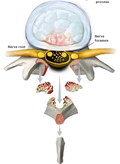
Conditions Treated with Laminectomy
Spinal Stenosis
Herniated Disc
Estimated Length of Stay in Hospital
The usual length of stay for a lumbar discectomy is 1-2 days depending on the patient’s medical condition and length of surgery. This may also be done on an outpatient basis as deemed by the surgeon.
Lumbar Discectomy (Microdiscectomy)
Overview
Lumbar Discectomy is removal of a disc in a specific level in the lumbar spine. By using a microscope and other instruments, surgeons are able to minimize the incision size which will increase healing time. Once the disc is removed, the nerves are no longer compressed or under pressure and can relieve the pain and numbness felt by the patients.
Procedure
After creating a small incision directly over the herniated disc, the surgeon creates a small window in the lamina (the bone covering the spinal canal). The pinched nerve root and the herniated disc can be seen through this opening.
Spinal Nerve Moved
The surgeon uses a nerve retractor to gently move the spinal nerve away from the herniated disc.
Herniation Removed
The herniated portion of the disc is removed, eliminating pressure on the nerve root. Only the
damaged portion of the disc is removed, leaving any healthy disc material to perform its function as a cushion between the vertebrae.
End of Procedure
The tools are removed, and the spinal nerve returns to its normal position. The incision is closed.
Conditions Treated with Lumbar Discectomy
Herniated Disc
Estimated Length of Stay in Hospital
The usual length of stay for a lumbar discectomy is 1-2 days depending on the patient’s medical condition and length of surgery. This may also be done on an outpatient basis as deemed by the surgeon.
Lumbar Interbody Fusion
Overview
Lumbar Fusion is a procedure where 2 or more vertebrae are fused together using bone graft and implants, rods, and screws which will eventually lead to one solid bone. The advanced technologies used can help eliminate pain by stopping abnormal movement and stabilize the spine. There are different approaches (anterior, lateral, and posterior) that are used separately or combined based on the anatomy of the disease and individualized patient evaluation.
Anterior Lumbar Interbody Fusion (ALIF)
Procedure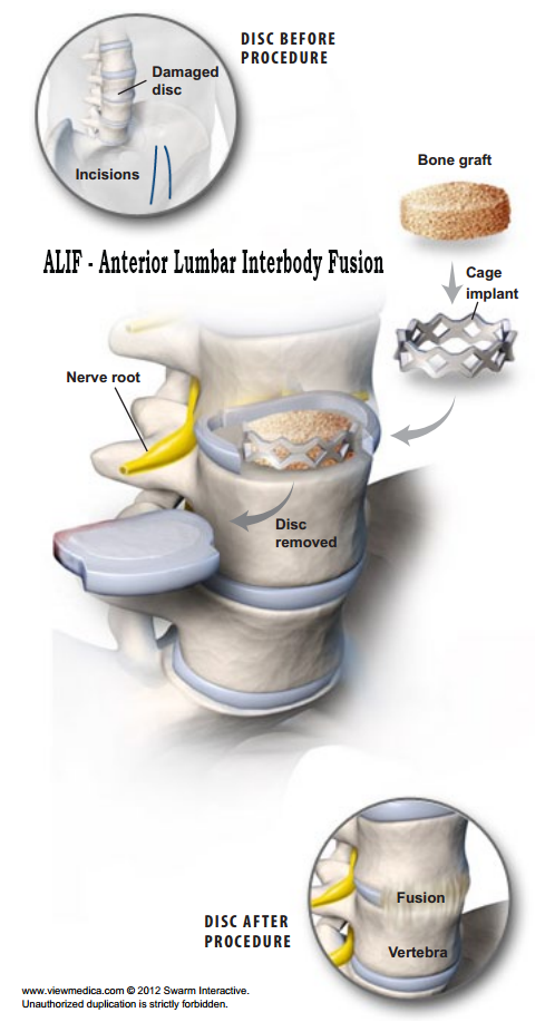
The procedure is performed through a three- to five-inch incision on the stomach. Two common approaches are over the center of the stomach or slightly to the side.
Disc Removed
The damaged disc is partially removed. Some of the disc wall is left behind to help contain the bone graft material.
Implantation
A metal cage implant filled with bone graft is placed in the empty disc space. This realigns the vertebral bones, lifting pressure from pinched nerve roots.
Vertebrae Secured
In some patients, this will be enough to secure the vertebrae. For others, the surgeon may need to implant a series of screws and rods along the back of the spine for additional support.
End of Procedure
Over time, the bone graft will grow through and around the implants, forming a bone bridge that connects the vertebra above and below. This solid bone bridge is called a fusion.
XLIF Lateral Lumbar Interbody Fusion
Procedure
Unlike traditional back surgery, XLIF® is performed through the patient’s side. By entering this way, major muscles of the back are avoided. This minimally-invasive procedure is generally used to treat leg or back pain
caused by degenerative disc disease. It can be performed on an outpatient basis.
Accessing the Spine
The surgeon creates two small incisions in the patient’s side. These incisions are much smaller than those used in traditional back surgery. A probe is inserted through one incision. The second incision is used to help
guide the surgical instruments.
Avoiding Nerves
The surgeon uses the probe to stimulate and detect nerves along the side of the spine. When a nerve is found it can be avoided and left undamaged. Fluoroscopic x-ray images also are used to guide the probe to the proper position on the spine.
Dilation Tubes Inserted
A series of dilation tubes are slid over the probe to create a larger opening. Then, a retraction device is used to move aside muscle tissue and gain access to the spine.
Disc Removed
The surgeon operates through the channel created by the retractor. The damaged disc is removed.
Implant Inserted
An implant filled with bone graft is placed into the empty disc space, realigning the vertebral bones. This also lifts pressure from pinched nerve roots. Bone Morphogenetic Protein (BMP) may also be used to encourage bone growth and a strong fusion.
End of Procedure
The morselized bone graft will grow through and around the implant, forming a bone bridge that connects the vertebral bodies above and below. This solid bone bridge is called a fusion.
Conditions Treated by Lumbar Interbody Fusion
Degenerative Disc Disease
Herniated Disc
Kyphosis
Scoliosis
Spinal Stenosis
Spinal Tumor
Vertebral Fracture
Estimated Length of Stay in Hospital
Usual length of stay is 5-10 days in the hospital depending on the amount of levels fused and patient’s other medical conditions. A brace will worn by the patients post-surgery for a time determined by your surgeon.
Kyphoplasty
Overview
Kyphoplasty is a procedure to stabilize vertebral fractures. A device is used to open the space and a cement-like material is implanted to support and stabilize the damaged vertebrae.
Procedure
Through a half-inch incision, small instruments are placed into the fractured vertebral body to create a working channel.
IBT Inserted
The KyphX® Inflatable Bone Tamp (IBT) is then placed into the fracture.
Cavity Created
The device is carefully inflated, creating a cavity inside the vertebral body.
Balloon Deflated
The balloon is deflated, leaving a cavity in the vertebral body.
Fracture Stabilized
The cavity is filled with bone cement to stabilize the fracture. Once filled, the incision is closed.
End of Procedure
With the process completed, an “internal cast” is now in place. This stabilizes the vertebral body and provides rapid mobility and pain relief. It also restores vertebral body height, reducing spinal deformity.
Conditions Treated with Kyphoplasty
Kyphosis
Osteoporosis
Estimated Length of Stay in Hospital
Usually this is done as an outpatient procedure although there may be circumstances that would lead to a 1-3 night stay based on patient’s medical condition.
Spinal Cord Stimulator
Overview
Spinal Cord Stimulator Insertion- A spinal cord stimulator is a device used to send electrical currents to the nerves in the spine to interfere with the nerve impulses that feel pain. This procedure is set up in two phases where a trial is used to ensure it works appropriately and adequately. Once the trial is over, a permanent stimulator is inserted. These are most commonly used for chronic pain patients.
Procedure
Spinal cord stimulation (also called SCS) uses electrical impulses to relieve chronic pain of the back, arms and legs. It is believed that electrical pulses prevent pain signals from being received by the brain. SCS candidates
include people who suffer from neuropathic pain and for whom conservative treatments have failed.
Trial Implantation
The injection site is anesthetized. One or more insulated wire leads are inserted through an epidural needle or through a small incision into the space surrounding the spinal cord, called the epidural space.
Find the Right Location
Electrodes at the end of the lead produce electrical pulses that stimulate the nerves, blocking pain signals. The patient gives feedback to help the physician determine where to place the stimulators to best block the
patient’s pain. The leads are connected to an external trial stimulator, which will be used for approximately one week to determine if SCS will help the patient.
Determine Effectiveness
If the patient and the physician determine that the amount of pain relief is acceptable, the system may be permanently implanted. At the end of the trial implantation, the leads are removed.
Permanent Implantation
The permanent implantation may be performed while the patient is under sedation or general anesthesia. First, one or more permanent leads are inserted through an epidural needle or a small incision into the predetermined location in the epidural space.
Generator Implantation
Next, a small incision is created, and the implantable pulse generator (IPG) battery is positioned beneath the skin. It is most often implanted in the buttocks or the abdomen. The leads are then connected to the IPG battery.
End of Procedure
The implant’s electrical pulses are programmed with an external wireless programmer. The patient can use the programmer to turn the system on or off, adjust the stimulation power level and switch between different programs.
After SCS Implantation
After surgery, patients may experience mild discomfort and swelling at the incision sites for several days.
Conditions treated by Spinal Cord Stimulator
Chronic Pain
Estimated Length of Stay in Hospital
This procedure is usually done as an outpatient going home the same day. There may be times that a patient will stay overnight based on the patient’s medical conditions or special circumstances.
